The Histopathology Laboratory at St. James’s Hospital is one of the largest histology laboratories in the Republic of Ireland. It provides a full range of services to the hospital and many other external healthcare providers nationwide, including other hospitals, GPs, and dentists. St. James’s Hospital is one of the eight designated cancer centres in Ireland and the laboratory handled nearly 30,000 requests in 2024.
In 2025, we introduced four new pieces of equipment that have improved how we work, speeding up sample processing and helping us deliver results faster, supporting better patient care.
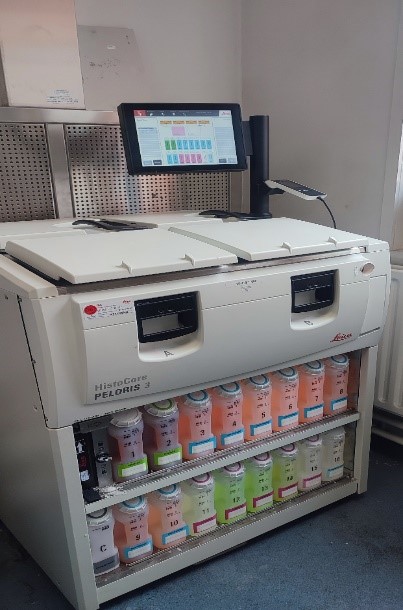
The Leica HistoCore Peloris 3 is a high-capacity tissue processor that can run two separate programmes overnight, allowing us to generate high quality samples. The new system has improved our workflow and reduced delays. As a result, patients benefit from faster, high-quality tissue processing and quicker turnaround times for results.
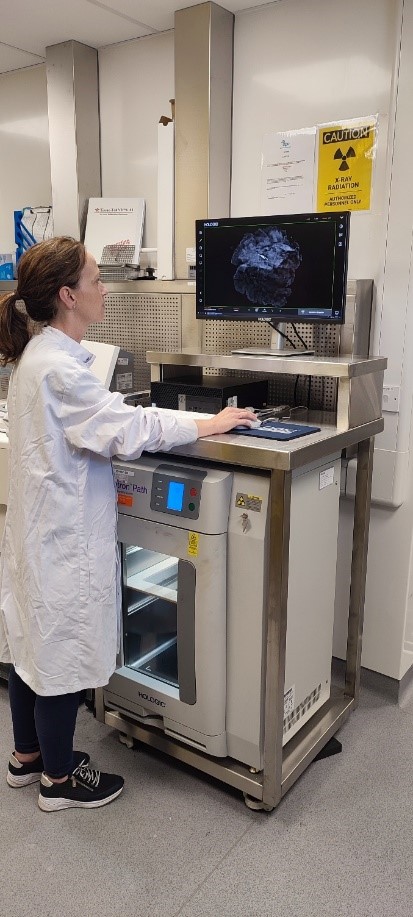
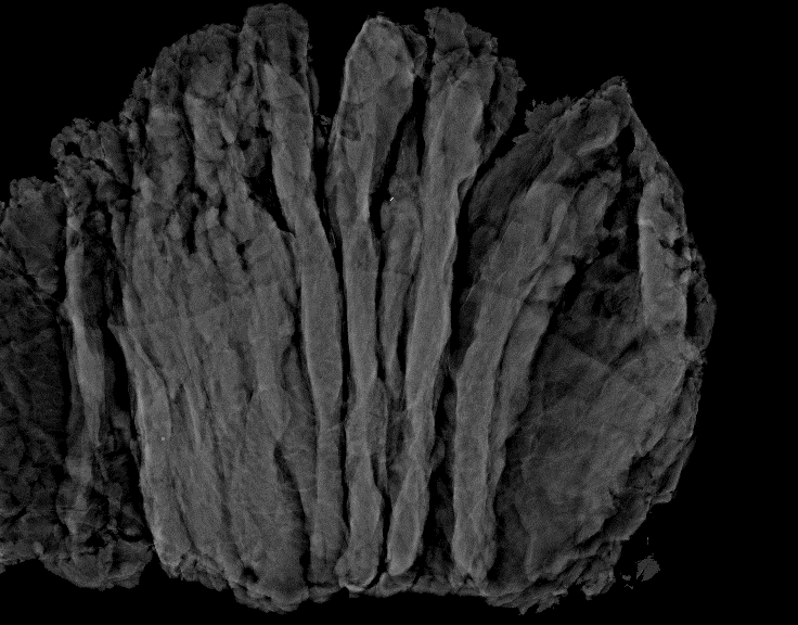
At St James’s Hospital, we use an X-ray machine called the Hologic Faxitron to take detailed images of breast tissue samples. These images help our team find tiny markers (called “clip sites”) that show where a biopsy was previously taken. The images guide staff, helping them focus on the most important areas of tissue for sample preparation.
The system also helps detect tiny calcium deposits in breast specimens, which can support the diagnosis of conditions like DCIS (ductal carcinoma in situ – a type of early-stage breast cancer). This means patients benefit from more accurate testing and fewer unnecessary samples being taken from large tissue specimens.
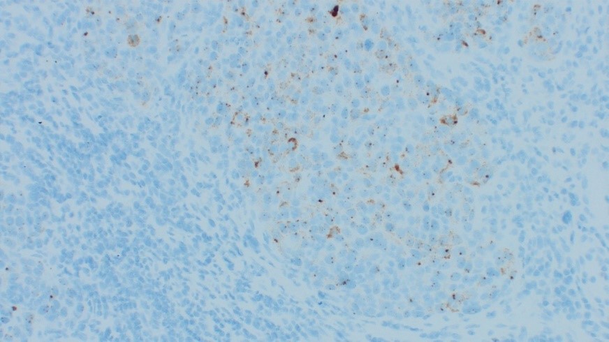
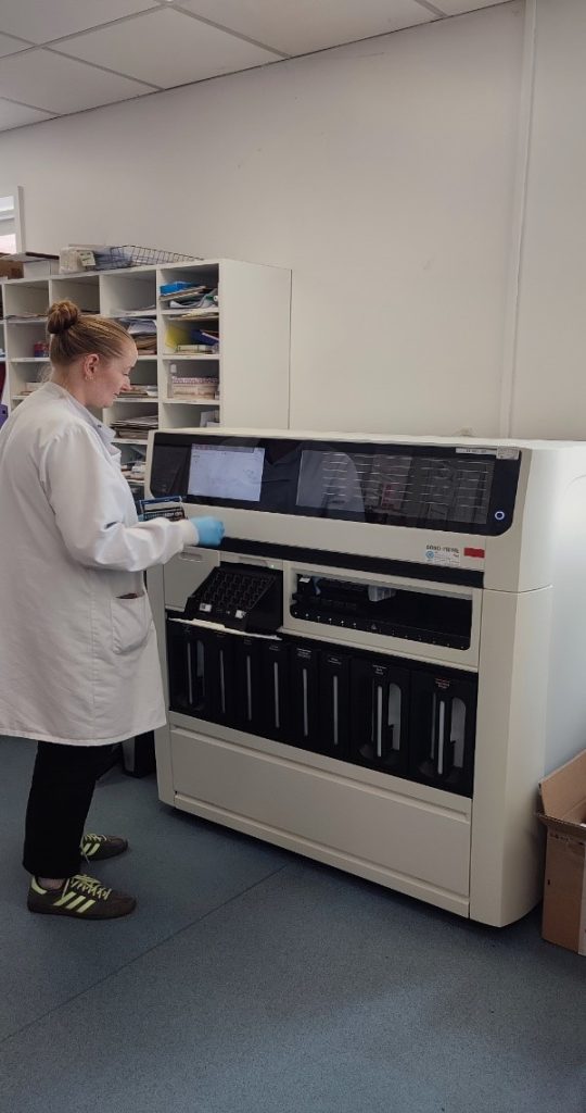
At St James’s Hospital, we use a machine called the Leica BOND-PRIME to carry out a test called immunohistochemistry (IHC). This test looks at tissue samples to help medical teams to understand what kind of specific illness a patient may have and how serious it is. In many cases, the results from this test help doctors decide which treatment will work best. The new machine works quickly and automatically, which means we can test more samples in less time. This helps us give faster results, supporting better patient care.
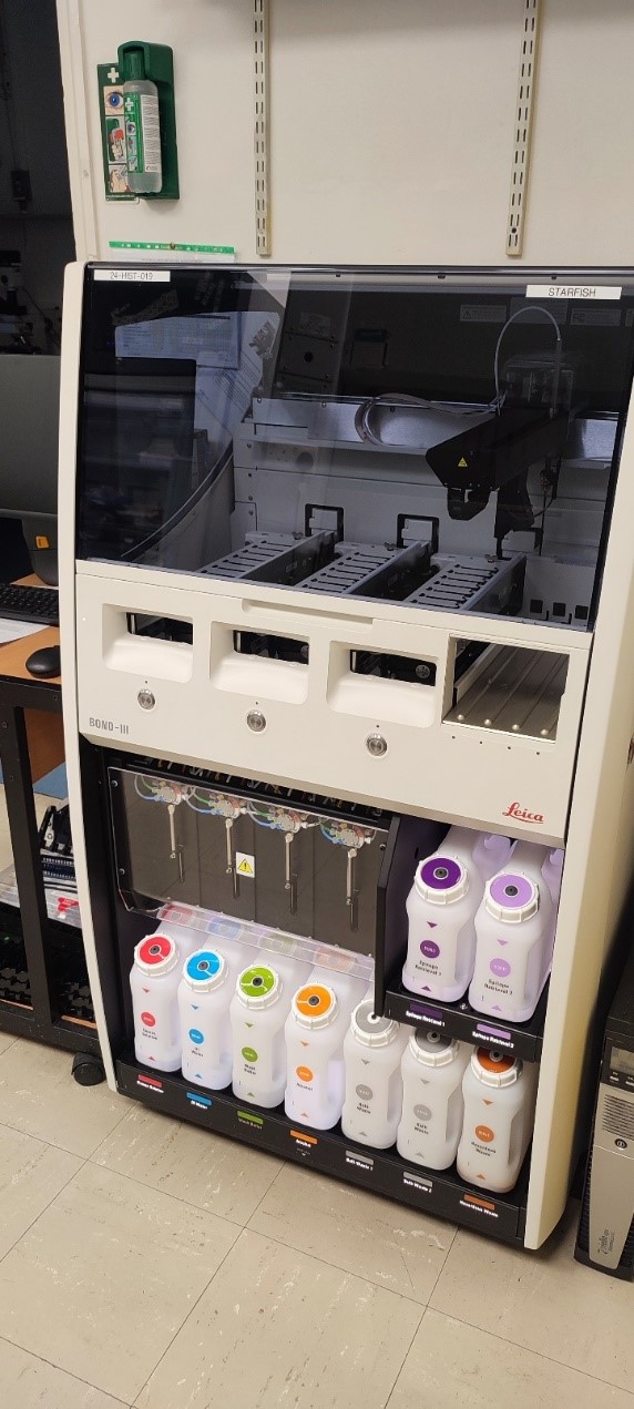
The Leica BOND-III analyser carries out advanced tests that help guide treatment decisions.
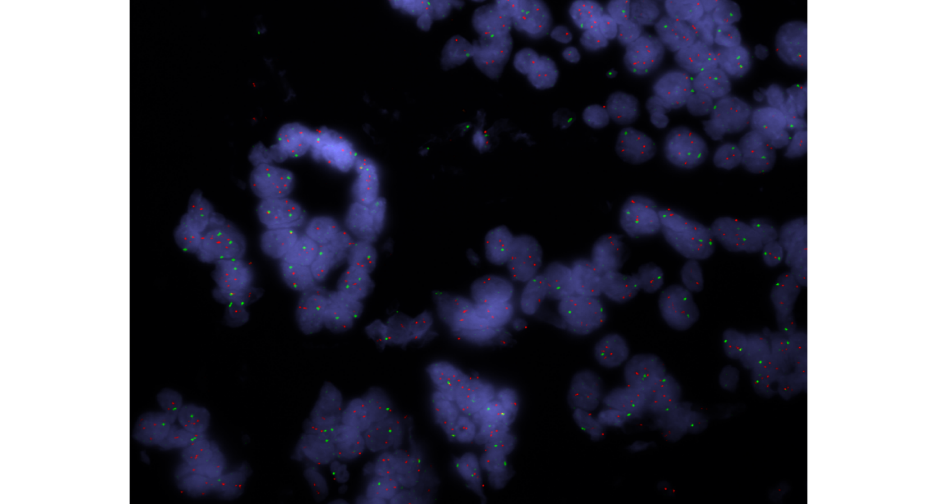
- HER2 FISH testing (Fluorescence in situ Hybridisation – a laboratory procedure used on breast cancer tissue to determine if the cancer cells have an amplified or extra number of HER2 genes) is used to support cancer diagnosis and treatment planning. With the new equipment, this test now runs automatically overnight, making the process faster and reducing delays. This gives our medical scientists more time to focus on reviewing and reporting results, helping us improve turnaround times.
- HPV RNAscope testing (a test used to detect the presence of human papillomavirus HPV within a patient’s tissue sample) is used for patients diagnosed with squamous cell carcinoma (SCC), which is a type of skin cancer. This test helps doctors understand the likely course of the disease and choose the best treatment for the patient.
By using this innovative technology, we’re able to offer reliable, efficient, and high-quality testing to support better care for our patients.